
13-jan-2024
Bowel atresia is a congenital condition in which the small intestine does not form correctly, leading to an obstruction in the intestines. It is one of the most common gastrointestinal birth defects, affecting 1 in 6,000 live births. There are several types of bowel atresia, including jejunal atresia, ileal atresia, and gastroschisis. These conditions can cause serious medical problems and require immediate treatment.
What is bowel atresia? A bowel atresia is a condition in which the small intestine does not form correctly, leading to an obstruction in the intestines. It can lead to other complications like malabsorption and infection, which could cause significant health problems and even death. There are several types of bowel atresia including jejunal atresia, ileal atresia, and gastroschisis.
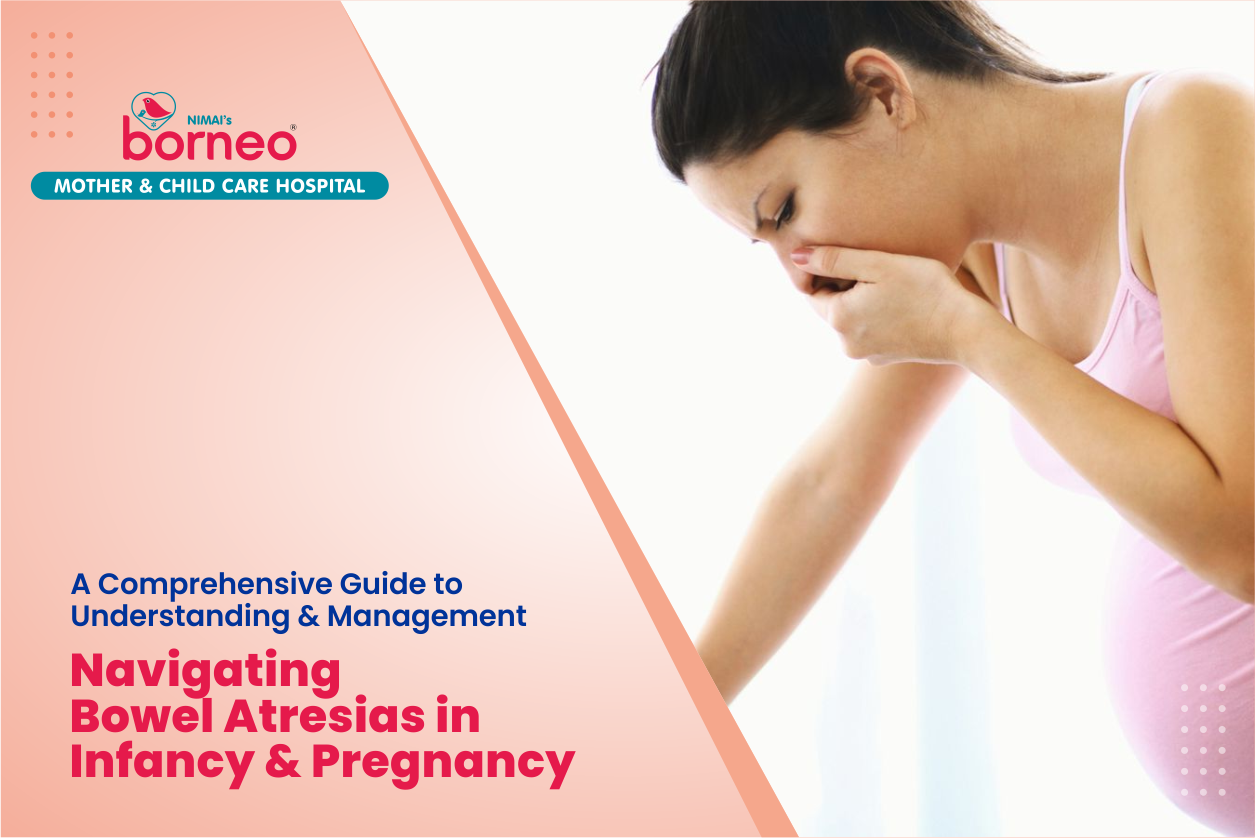
There are several potential symptoms of bowel atresia including fever, lochia (blood loss), vomiting, diarrhea, weight loss, and abdominal pain. Bowel atresia may be diagnosed before birth through a combination of ultrasound and fetal echocardiogram. In other cases, the diagnosis is made after birth through abdominal or chest x-ray.
A variety of factors can cause bowel atresia including genetic conditions such as Prader-Willi syndrome and Ewing's sarcoma; environmental toxins like Listeria monocytogenes; and intrauterine infections that cause the womb to contract abnormally.
Bowel atresias are a type of congenital malformation in which there is an obstruction of the intestine due to a narrowing or complete closure of the lumen. This condition can lead to serious health complications in infants, including vomiting, abdominal distension, and failure to gain weight. Diagnosis and treatment of bowel atresias require specialized pediatric surgery. The first step in diagnosing bowel atresias is to establish the clinical history of the infant, including date of birth and weight. Next, a physical examination should be conducted to check for signs of abdominal distension or other signs that characterize bowel atresias. To perform this examination, the physician will use a stethoscope to listen to the abdomen and then assess whether there is any fluid with audible peristalsis present. If so, one must rule out obstruction due to an intestinal malrotation or volvulus (a twisting motion) that may have been caused by abdominal trauma. A radiograph of the abdomen should also be taken to rule out any signs of obstruction. Diagnosis and management of bowel atresia during pregnancy includes prenatal diagnosis, antenatal management strategies, antenatal care, and postnatal care. Prenatal diagnosis of bowel atresia includes abdominal ultrasound scan, fetal biophysical profile (FBP), and an abdominal x-ray. Ultrasound scan is recommended to confirm if the anus is patent or non-patent (closed). Biophysical profile can be used in order to differentiate between oligohydramnios and hydramnios. A postnatal diagnosis of bowel atresia is a biopsy of the rectum, which confirms the diagnosis. antenatal management strategies for bowel atresia include conservative management, expectant management with oral ursodeoxycholic acid (UDCA), and prophylactic surgery. The most common surgical procedure used to treat bowel atresias is jejunal intussusception repair. This procedure involves resecting the affected segment of intestine and re-anastomosis it with healthy tissue. Other treatments may include gastrostomy tube placement and/or endoscopic balloon dilatation. With proper diagnosis and treatment, most infants with bowel atresias can go on to lead normal lives
Before birth, bowel atresias are diagnosed through ultrasound. Scans of the abdomen help to detect any anomalies in the organs of the intestines that may be associated with this condition. The size, shape and location of these anomalies will help doctors to identify which tract is affected by the atresia, as well as its severity. Depending on how severe a bowel atresia is, doctors will recommend either a planned vaginal delivery or cesarean section. If vaginal delivery is chosen, then it may require an expedited labor and cesarean section to deliver the first baby in case there are complications during the labor. A healthy bowel is a tube-like structure that carries food and waste from the stomach to the colon. This process begins in utero with the formation of a fetal anus, which closes around 4–5 months gestation. At about 6 weeks post conception, a small opening at the bottom of the rectum is formed and will eventually close off completely when it becomes complete in late pregnancy or shortly after birth.
Nimai Institute of Medical Sciences Private Limited (NIMS) was founded by its founder chairman Dr Santosh N. Madrewar, a renowned Pediatrician, who has designed an unique Mother and Child Health Concept, integrating modern medical management with Ayurvedic treatments and traditional Indian ethos making Pregnancy and Birthing a Joyous Celebrations and Child development focused on Physical, Mental, Social and Spiritual Well-being.
Chat Now